Simulations of Eye Disorders
This page shows simulations of visual symptoms caused by a variety of eye disorders, from simple refractive error, to localized eye problems, to neurological problems. Please note that these simulations are generally based on the author's interpretation of symptoms described by patients, as well as on knowledge of the disease processes themselves. There may be wide variations in an individual's own visual experiences with any disease process.
Please Note:
These sections are not intended to replace the professional examination and diagnosis by a physician, and they are presented here purely for informational purposes. All possible diagnoses and treatment options are not covered, and the information discussed should not be taken as a recommendation to self-diagnose and self-treat a condition. A misdiagnosed or improperly treated eye condition can result in a permanent loss of vision, or a permanent loss of function of the eye or visual system. In the case of any eye problem, seek medical attention promptly. This can include emergency room treatment, as well as treatment by a medical physician or eyecare provider.Refractive Errors

Normal Image
Refractive errors are optical abnormalities in the eye that lead to improper focusing of images onto the retina. Refractive errors are generally correctable by glasses, contact lenses, or refractive surgery. Thus, blurred vision due to refractive error alone does not cause an untreatable loss of vision (the eye is otherwise healthy). For more information on the optics of the eye and refractive error, go to Optics and Refractive Error.
This is a normal image of the Italian Gardens at Maymont Park, Richmond VA. Both the foreground and the far distance are clear. »
Nearsightedness

Refractive Error: Myopia
In this myopic (nearsighted view) of the gardens at Maymont Park, the foreground is clear but the midrange and distance become progressively blurrier.
Farsightedness

Refractive Error: Hyperopia
Both distance and near are blurred, but the distance is relatively clear compared to the foreground.
Astigmatism
Astigmatism is usually caused by the cornea not being perfectly round in all directions, leading to different parts of an image being blurred, depending upon the orientation of the astigmatism. In this image of Capitol Square in Richmond, Virginia, the astigmatism is oriented vertically, thus there is selective blurring of vertical lines as shown in the photo.

Refractive Error:Astigmatism (Uncorrected View)

Refractive Error:Astigmatism (Corrected View)
Cataract

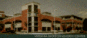
Normal View
Clear image of the Virginia Biotechnology Center in Richmond, Virginia.
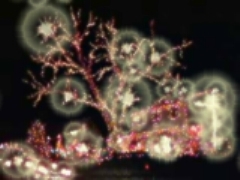
Nuclear Sclerotic Cataract
Nuclear Sclerotic Cataract
This image represents the view through a "nuclear sclerotic" type of cataract. Note that in addition to the image being blurred, it is also dim and less colorful than the normal view. The color blue is especially obscured by this common type of cataract.
Posterior Subcapsular Cataract

Normal View
An infamous decorated house on Asbury Ct. in Richmond, Virginia.
Cataract View
The "posterior subcapsular" type of cataract often affects younger individuals, and those with diabetes or on steroids. There may be severe glare with this type of cataract, with halos and starbursts being visible from point sources of light.
Glaucoma
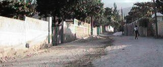
Normal View
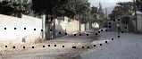
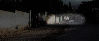
This is a normal view of a street with no loss of peripheral vision.
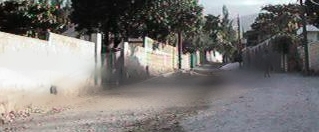
Glaucoma View
This demonstrates a common type of blind spot found early in glaucoma. If the eye is fixating down the road, the pedestrian on the right partially disappears. There is a reduction of sensitivity in the peripheral vision in the area shown within the dotted lines below.
Glaucoma, severe loss of vision
This shows a severe loss of visual field in advanced glaucoma, left eye. Only an island of central vision remains, with some field of vision toward the outside left as well. The blind spot cuts horizontally through the center of vision, an especially severe loss of vision.
Macular Degeneration
Normal View
Clear image of a building in this case with no macular degeneration.
Early "Wet" Type AMD
Early "Wet" Type View
This demonstrates distortion typical of early neovascular ("wet") macular degeneration, with distortion of the retina. Although distorted, the lettering is still readable.
(Other conditions causing swelling of the central retina may cause this visual distortion, such as cystoid macular edema, central serous choroidopathy, diabetic macular edema, among others.)
Severe "Wet" Type AMD
Severe "Wet" Type View
In this more severe case of "wet" macular degeneration,
the vision is distorted and also lost centrally.
Retinal Detachment

Normal view of Richmond skyline.

In the area of a retinal detachment, vision is lost.
At the margin of the detachment, the vision may be distorted.
Vitreous Hemorrhage

Normal view of a lake at Lewis Ginter Botanical Gardens in Richmond, Virginia.

Eye with strands of blood creating a veil over the vision. This represents a minor hemorrhage into the vitreous body of the eye. Sources could include diabetic retinopathy, retinal tears, vitreous detachment, and sickle cell retinopathy, among others.
Double Vision

Double vision looking down a road leading into Shockoe Slip, in downtown Richmond. In this case, the double vision is oriented horizontally (side by side). This could occur with dysfunction of certain nerves controlling eye movement, or with thyroid related orbital problems.
Color Blindness
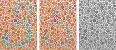
These images demonstrate how individuals with normal color vision, and abnormal color vision, might view this color vision test. Those with normal vision would see the number "3" as shown on the left. With mild color blindness (as is present in about 8% of males), the number may be misread as a "5", as shown in the middle. With total color blindness, no number or pattern is visible, as shown on the right (rare).
Ophthalmic Migraine

This is a depiction of the visual phenomenon experienced by many before a migraine headache. The area of jagged, zigzag lights are constantly in motion, flashing over a 15 to 30 minute time frame. The area involved often starts small near the center of the vision, then moves outward slowly. There is a scotoma, or a blink spot, in the area of the disturbance, which is seen in the same field of vision of both eyes. A migraine headache may, or may not, follow this aura.